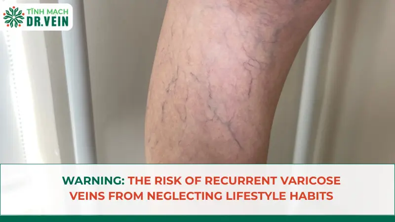
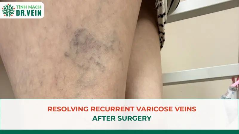
Recurrent varicose veins can cause venous clusters to continue appearing or developing over time if the damaged venous system is not comprehensively treated. When these manifestations persist for many years post-intervention, re-evaluating the venous system using Doppler ultrasound is a mandatory medical indication to determine the underlying cause and develop an appropriate treatment plan for recurrent varicose veins.
In recent years, Vietnamese medicine has increasingly asserted its position internationally with a team of highly specialized doctors in intensive treatment fields. During a combined business and leisure trip to Ho Chi Minh City, Ms. Arina (45 years old, Canadian nationality) chose the Dr. Vein medical system to examine her bulging veins that had persisted for years.
This trip helped her discover the root cause behind the increasingly prominent venous clusters on the back of her legs, 6 years after her previous intervention in Canada.
The silent progression of recurrent varicose veins
Approximately 6 years ago, Ms. Arina was diagnosed with venous disease and underwent Endovenous Laser Ablation (EVLA) for the great saphenous vein in Canada.
Following the treatment, the initial symptoms subsided. However, in the subsequent years, she began to notice prominent venous branches on the back of her lower legs. Initially presenting as small blue veins, they progressively dilated, became tortuous, and formed bulging clusters on the skin surface over time.
After 6 years, these venous clusters developed to a severe degree, leading the patient to mistakenly believe this was a natural progression of aging and that she had to accept living with the condition. Trusting in the internationally reaching Vietnamese medical sector, she decided to visit the Dr. Vein venous specialty clinic for a comprehensive reassessment.
Doppler ultrasound diagnoses the cause of recurrent varicose veins
At Dr. Vein, Dr. Le Kim Cao directly conducted the clinical examination and performed a lower extremity Doppler vascular ultrasound.
Clinical results indicated that the great saphenous vein had been completely occluded from the previous intervention. However, the small saphenous vein still exhibited severe valvular insufficiency with prolonged reflux. Simultaneously, the superficial venous branches on the back of the lower leg had severely dilated, forming distinct venous clusters. The pathology was classified as stage C2 according to the CEAP scale.
This result enabled the doctor to pinpoint the direct correlation between the residual small saphenous vein insufficiency and the recurrent varicose veins on the back of the leg over the past 6 years.
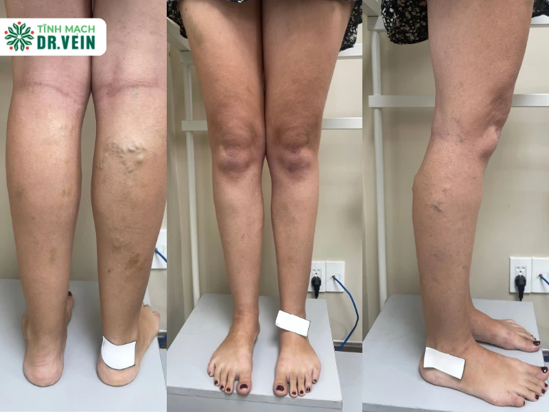
Medical explanation: Why do venous clusters continue to appear?
The medical record revealed that Ms. Arina had venous insufficiency in both the great and small saphenous veins. However, the previous intervention protocol in Canada only treated the great saphenous vein.
After the occlusion of the great saphenous vein, the insufficiency in the small saphenous vein persisted. The refluxing blood continuously exerted pressure on the superficial venous system at the back of the lower leg, weakening the vein walls and causing the venous branches in this area to dilate.
This process occurred silently. The initially small veins gradually developed into large, tortuous clusters under the skin. Therefore, Ms. Arina’s condition was not a superficial sequela but the consequence of the underlying insufficiency in the small saphenous vein not being completely eradicated. The scope of the previous treatment failed to cover the entire damaged venous system.
According to the Clinical Practice Guidelines of the European Society for Vascular Surgery (ESVS), re-evaluating the entire hemodynamic system using Doppler ultrasound is a mandatory step when treating recurrent varicose veins. Ms. Arina’s case clearly demonstrates the principle: When venous clusters reappear in a new location, a comprehensive survey is required rather than focusing solely on the previously treated segment.
Multi-method intervention protocol
For this clinical case of recurrent varicose veins, Dr. Le Kim Cao prescribed a combined treatment protocol:
- EVLA Intervention: Thermal energy from the laser completely ablates the insufficient small saphenous vein segment, permanently closing the pathogenic reflux source.
- Muller Phlebectomy: Direct dissection and removal of the severely dilated, tortuous venous clusters on the back of the lower leg through micro-incisions, ensuring maximum aesthetic results.
Given that the venous clusters had existed for 6 years with severe structural alterations, direct removal via the Muller method was necessary instead of waiting for the blood vessels to atrophy spontaneously after treating the reflux source. This combination provides a dual therapeutic effect: resolving the root cause while thoroughly eliminating the surface clinical manifestations.

Thanks to the accurate diagnosis of the underlying cause, Ms. Arina was relieved of the psychological burden of having to live with the disease. This clinical case not only affirms the importance of Doppler ultrasound in managing recurrent varicose veins but also demonstrates the professional capacity of the Vietnamese healthcare system in successfully receiving and treating complex cases for international patients.